

Eucharistic Miracles:
God Under the Microscope
“The God of the Bible is also the God of the genome. God can be found in the cathedral or in the laboratory.”
— Francis Collins (1950-)
Head of the Human Genome Project, Director of NIH
By Chris and the Editorial Staff

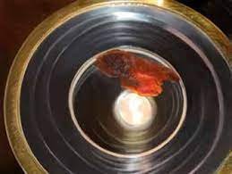
The Communion host from Legnica, Poland in 2014, found to contain human heart muscle in a study by Pomeranian Medical University (Poland).
5 Investigations, 3 Continents, 40+ Years
During the Last Supper, Jesus told his 12 apostles, “Take and eat; this is my body. Drink… this is my blood” (Matthew 26:26–28). Catholic teachings hold that the bread and wine that priests bless during Mass transform into the body and blood of Jesus through transubstantiation. Under the guise of bread and wine, Jesus’ body and soul constitute the “Eucharist,” which Catholics receive and consume during Mass.
Ordinarily, the physical appearance and chemical composition of the bread and wine remain unchanged despite Christ’s presence in the Eucharist. However, hundreds of Eucharistic phenomena, where consecrated bread physically transforms into human flesh or blood, have been reported over the last 2,000 years.
With advancements in technology, the Catholic Church is now able to thoroughly investigate these events, enlisting the help of scientists using modern scientific techniques. This article focuses on five investigations where these details have been published, from 1971 to 2014.
Amazingly, all five scientific investigations of these Eucharistic phenomena identified heart tissue under the microscope. Two characterized the samples as type AB, a rare blood type present in 6% of humans. The map below shows the location and year of each sample (yes, the Lanciano sample is ancient).

Miracle or Fraud?
Because the Church investigates various claims of miracles, many have been exposed as hoaxes. For example, a church custodian was accused of using his own blood to simulate a bleeding statue after the DNA of the blood on the statue was found to match his own DNA. Household materials such as red paint are easily identifiable through chemical analyses.
In contrast, the five Eucharistic phenomena that we will discuss all stood up to extensive scientific investigations. Scientists scrutinized these tissues under the microscope and used immunohistochemical tests, chromatography, and other lab tests to determine their composition and characteristics. Many of these scientific analyses are publicly available, albeit in foreign-language publications, and several of these reports are linked from this page.
Almost every scientific investigation of the real world is going to have some limitations, but what makes these phenomena compelling is the similar findings across events separated by thousands of miles—and, in one case, over a thousand years. We will first focus on each case under the microscope before we zoom out to take a big-picture look at what makes these phenomena so intriguing together as a whole.

The Communion host at Sokolka, where the red tissue is joined to a host fragment (top left). The underlying surface is a linen corporal with an embroidered red cross.

The monstrance displaying the host at St. Anthony of Padua Church in Sokolka
Sokolka, Poland (2008)
The first Eucharistic phenomenon we will discuss occurred at St. Anthony of Padua Church in Sokolka, Poland. On October 12, 2008, a priest placed a host (a piece of consecrated bread) in a container of water after it had fallen to the ground. Consecrated hosts that become dirtied are usually dissolved in this way so that they can be poured into a sacrarium for disposal. Sister Julia Dubowska, the parish sacristan, placed the container in the sacristy’s safe. One week later, she was astonished to find in the container a red substance connected to a partially dissolved host, and she quickly informed the other priests.
After 18 days of submersion in water, the tissue and the associated host were moved to a linen corporal and left to dry. In January 2009, the archbishop asked two anatomical pathologists from the Medical University of Bialystok to examine the tissue. Professor Maria Elżbieta Sobaniec-Łotowska and Professor Stanislaw Sulkowski were both highly respected pathologists in their university who had each published dozens of research articles in peer-reviewed journals. Sobaniec-Łotowska took a small sample of the red portion, along with its connection to the host, and gave half of it to Sulkowski for microscopic analysis. He was not told of its origins at first so that he could independently analyze the tissue without prior biases. The professors each came to the same conclusion after inspecting the tissue with both light and electron microscopy: The samples were heart muscle.
The Polish newspaper Nasz Dziennik interviewed Sobaniec-Łotowska and Sulkowski in December 2009. The following is an excerpt from that interview:
Sulkowski: If we put the Communion wafer in the water, in the normal course of events it should dissolve in a short time. In this case, however, part of the Communion, for some incomprehensible reason, did not dissolve. Moreover, what is even more incomprehensible—the tissue that appeared on the Communion was tightly connected to it—infiltrating the substrate on which it was formed. Take my word for it that even if someone had intended to manipulate it, he would not have been able to connect the two structures so inseparably.
Sulkowski found two things astounding about this sample. First, the Communion wafer, which contains only flour and water, did not decompose after 18 days of submersion in water. Second, the bread and cardiac muscle tissues were intricately interwoven in a way that would be impossible to accomplish through human manipulation.
Sobaniec-Łotowska: This remarkable phenomenon of the intermingling of the Communion and the fibers of the heart muscle observed in both light microscopes and transmission electron microscopy also demonstrates to me that there could be no human interference here. In addition, please note another unusual phenomenon. The Communion stayed in the water for a long time, and then even longer on the corporal. Thus, the tissue that appeared in the Communion should have undergone a process of autolysis [a type of necrosis or tissue death]. Examining the collected material, we found no such changes. I think that at the current stage of development of knowledge, we are not able to explain the studied phenomenon solely based on natural science.
Transmission electron microscopy can be used to visualize incredibly small details, including viral particles and atoms. After using this exquisitely sensitive tool, Sobaniec-Łotowska agreed with Sulkowski’s assessment of the interwoven fibers. This integration could not have been achieved by any human craft. She also affirmed that the cardiac tissue should have decomposed in water, yet it remained intact without any signs of degradation.
Because of these astonishing findings, Sobaniec-Łotowska and Sulkowski were formally reprimanded by their university and accused of carrying out “illegal” and “disloyal” investigations that incorporated the “emotional” aspect of their Catholic faith (Serafini chapter 4). A tabloid magazine article speculated that the red substance might have been bacterial contamination with Serratia marcescens, even though these rod-shaped bacteria look nothing like heart tissue under the microscope. The president of the Polish Rationalist Association even initiated a frivolous lawsuit calling for a criminal investigation for murder since the heart tissue must have come from someone.
Sulkowski defended what he did (Serafini chapter 4):
We have the duty to investigate every scientific problem… Just as a doctor cannot refuse to care for a patient, likewise, we have the duty to research every scientific problem, according to the guidelines of the Polish Academy of Sciences.
Yet their report led to more questions than answers. Where did the heart muscle come from? Why didn’t the heart tissue decompose after 18 days in water? How did the muscle and host become so intertwined that two experts independently concluded that a human could not have fabricated it? Science cannot currently offer satisfactory answers to these questions.
It is natural then to consider fraud. Only two people had keys to the safe with the transformed host, but let’s imagine that someone got access and wished to publicize a miracle to garner attention. It’s difficult to envision such a person going to the trouble—if they even had the ability—to fabricate a piece of heart tissue interwoven with bread in the anticipation that it would later be examined under an electron microscope.
Reporting these scientifically inexplicable findings only harmed their professional reputations at their university, so Sobaniec-Łotowska and Sulkowski lack any obvious motive for colluding or falsifying their strange results when they were already respected for publishing traditional journal articles. On the contrary, their rigorous approach convinced them to stand by their objective findings despite the surrounding controversy. Their results highlight both the usefulness of science in confirming a tissue’s identity and the limits of our current knowledge of science to explain everything. If one believes, as the Church does, that this event was a Eucharistic miracle, these mystifying findings are part of the miracle.

Professor Maria Sobaniec-Łotowska
Medical University of Bialystok
Research Gate (130+ publications)

Dr. Barbara Engel, a cardiologist on the Legnica ecclesiastical committee
Legnica, Poland (2013)
Five years later and 400 miles away, another Eucharistic phenomenon occurred in Legnica, Poland. During the first Christmas morning Mass of 2013 at Sanctuary of St. Hyacinth, a consecrated host fell on the ground. The priest placed the host in a chalice filled with tap water and left it in the tabernacle to dissolve.
On January 5, 2014, Father Andrzej Ziombra went to check on the host. He reported his discovery:
Immediately we noticed that the Host had not dissolved, and that a red spot covering a fifth of Its surface appeared. We decided to inform the Bishop, who established a special theological scientific commission to analyze the event.
Over the next two weeks, the rest of the host dissolved in the water and only the red portion remained (see pictures below documenting the changes over time). On February 10, this red material was placed onto a corporal cloth and left to dry. Fifteen specimens were taken from this red tissue in the presence of witnesses, with each step of the sampling process photographed. Control samples were also prepared from both non-consecrated bread and consecrated hosts from the same production batch as the host that had developed the red tissue. The Institutes of Forensic Medicine at two Polish universities, Wrocław Medical University and Pomeranian Medical University, studied these samples. Wrocław Medical University ruled out the presence of bacteria and fungus and deemed the turning of the bread host into tissue scientifically inexplicable (Serafini 101). A lab report from Pomeranian Medical University identified the tissue as human heart muscle.
Dr. Barbara Engel, head of the cardiology department at the Provincial Specialist Hospital in Legnica, was a member of the ecclesiastical committee overseeing the investigations. She discussed the findings in an interview with Olivier Bault from the French newspaper Présent in December 2016.
Bault: I read that two research organizations have studied this host.
Engel: In fact, not all the scientists and organizations that initially participated in this study agreed to give an opinion. Many withdrew, citing various reasons after learning the origin of this sample. In the end, two organizations issued a scientific opinion. These are the Institute of Forensic Medicine in Wrocław and the Institute of Forensic Medicine in Szczecin.
Bault: Did those who withdrew question the nature of this matter?
Engel: No, they agreed that it was myocardial [heart muscle] tissue, but when told where this fragment came from they refused to write an opinion.
The reluctance of scientists and institutions to publicize their findings underscores why the Eucharistic phenomena are not discussed more widely among scientific circles. The Medical University of Bialystok’s reprimand of Sobaniec-Łotowska and Sulkowski in Sokolka just five years earlier probably did not inspire confidence that yet another report of heart muscle appearing on bread would be well received by the scientific community. Many scientists may be reluctant to be associated with any event described as miraculous. Even if their slides showed heart tissue, they may have had doubts about where it came from.
Bault: We could read in the newspaper Gazeta Wyborcza, and therefore in an article written by people who were not necessarily believers, that the method of taking the samples had not fulfilled the criteria usually accepted for this kind of analysis, particularly in forensic medicine. Is it true?
Engel: If that's what we read, the author of this article was misinformed. He would have had to find out how the material was taken, which obviously was not the case. The samples were taken by the Institute of Forensic Medicine in Wrocław. The whole procedure was recorded, documented, and described. The scientists, when taking the samples, filmed and photographed everything. The collection of samples for analysis was done in an absolutely professional manner. Then, the procedures carried out in the laboratories of this institute were in turn documented. Each sample has been properly isolated, described, packaged, transported, etc. In scientific circles, the procedure followed did not raise any questions.
Critics also brought up concerns about possible contamination with Serratia marcescens, a type of bacteria that produces a vivid red pigment, but this theory does nothing to explain how cardiac tissue was found under the microscope.
We now have many scientific tools at our disposal that can shed light on mysterious events. Just as the Catholic Church has welcomed scientific investigation of religious phenomena, perhaps the scientific community should be more open to investigating these events to the extent possible. Scientific progress is jeopardized when scientists do not report objective findings about religious phenomena because they are afraid of tarnishing their professional reputation or standing in the scientific community.
Bault: And so, to recap, from your point of view there is no doubt that we are dealing here with a Eucharistic miracle?
Engel: For me, as for everyone who has seen this Host up close, there is no doubt that it is a miracle. That said, medicine is an exact science but theology, contrary to appearances, is also an exact science. Here, the bishop tells us, as the Church teaches, that to say that there is a Eucharistic miracle, this transformation of the Host must be accompanied by extraordinary phenomena. This is why we speak here of an “event having the characteristics of a Eucharistic miracle.” This event is now observed. It is only if other inexplicable events occur around this Host that we will speak in the future of a “Eucharistic miracle.” So much for the theological side. But as far as I am concerned, I am not a theologian and it is indeed a miracle.
Bault: So I come back to my question: what has changed in your life?
Engel: It is difficult for me to answer this question, but I can say without hesitation that I received a huge injection of faith. This has been accompanied by many graces in my daily life.
This investigation demonstrates how science can coexist with religion and even nourish faith.
Translated excerpt of the report from Pomeranian Medical University

The monstrance containing the tissue at St. Hyacinth’s Church

After two weeks, the host almost completely dissolved while the red tissue remained.

The tissue was removed from the water and placed on a cloth for later analysis.

The red color appeared on the host while it was dissolving in water.

The red portion detached from the host a few days later.

Fragment of cardiac muscle fiber found in the sample
Tixtla, Mexico (2006)
Poland is not the only country that has witnessed Eucharistic miracles. Another transformation took place across the Atlantic Ocean in Tixtla, Mexico. In 2006, Father Leopoldo Roque, pastor of the Parish of Saint Martin of Tours, invited Father Raymundo Reyna Esteban, known affectionately as Father Rayito, to lead a spiritual retreat for his parishioners.
The 600 retreat attendants filed into the auditorium to celebrate the Sunday Mass on October 22, 2006. During the Communion distribution, Sister Arely Marroquín noticed one of the hosts exuding a reddish substance that looked like fresh blood. The Eucharistic host was reserved for later study.

The bleeding host of Tixtla shortly after the event on October 22, 2006
In the weeks following the event, Monsignor Alejo Zavala Castro, the bishop of the local diocese of Chilpancingo, set up an inquiry committee that collected the sworn testimonies of witnesses. Three years later, Dr. Ricardo Castañón Gómez, a clinical psychologist who was interested in studying Eucharistic phenomena, contacted Bishop Castro. Dr. Castañón had founded a company, Grupo Internacional para la Paz, that recorded the sampling of relics and forwarded them to a wide network of laboratories in a “clear, unbroken and documented chain of custody” under blinded conditions (Serafini chapter 2). He was appointed to begin investigations of the Tixtla host in 2009 and collected samples that were sent to multiple forensic labs.
Dr. Orlando Rodas Pernillo and Dr. Elisa Hernández de Rodas, specialists in Surgical, Oncological, Gastrointestinal Pathology and Cytology at Patología Médica, reported the following:
The architectural fibers showed bifurcations, which can be seen in cardiac muscle. The structure of the tissue was consistent with muscle tissue from the heart, but definitive microscopic identification was not possible because of the decomposition of the sample. (Immunohistochemical tests by another lab confirmed that this was cardiac muscle.)
Several kinds of cells were visualized: fat cells, red blood cells, and white blood cells. The microscope slide even showed a macrophage, a type of white blood cell, in the middle of ingesting fatty debris.
The Gene-Ex Genetics Laboratory in Bolivia found the following:
An immunoassay for the detection of human hemoglobin was positive. This test was specific for the human version of hemoglobin, a protein in red blood cells, confirming that the blood was human.
The blood was type AB negative. The AB negative blood type is the rarest blood type in the world, comprising less than 1% of the population.

Longitudinal fibers with bifurcations, typically seen in cardiac muscle.
Dr. Eduardo Sánchez Lazo, a professor of Legal and Forensic Medicine at the National Autonomous University of Mexico, also studied a sample and signed a statement with the following conclusions:
“The immunological study shows that the blood is of a human, and the immunohistochemical test allows us to state with all objectivity and certainty that it belongs to blood group AB.” This confirmed the findings from the Gene-Ex laboratory.
“After immunohistochemical tests, it has been determined that the tissue under study corresponds to the heart due to its macroscopic characteristics, in addition to showing the aforementioned cytochemical results.”
“The sample presents … an outflow of blood from its interior to its periphery, that is, the blood comes from the interior to the exterior… The possibility that the bleeding comes from the outside inwards is ruled out.” To evaluate for signs of fraud, Bishop Castro had requested that the doctor study whether the blood had been added to the host from the outside. Dr. Lazo confirmed that there was no external source of blood.
This investigation engaged laboratories and scientists in multiple countries who worked independently. They were not told where the specimen came from or what the other laboratories had found. Laboratories used diverse, complementary techniques—microscopic evaluation and an array of immunological and biochemical tests—to analyze the sample and ultimately arrived at the same conclusion: This was heart muscle and type AB human blood.
The extensive investigations of the Tixtla host confirmed that this was truly biological tissue, containing muscle, fat cells, red blood cells and white blood cells. Furthermore, the human blood came from the interior of the host and spread outward instead of originating from the outside, which calls to mind Sobaniec-Łotowska’s assertion that no human interference could have created the intermingled bread and heart tissue in Sokolka. Underlying these findings is the same tissue that was present at both Eucharistic events in Poland. Heart muscle is inexplicably found in every Eucharistic phenomenon discussed on this page.

Dr. Frederick Zugibe, chief medical examiner of Rockland County, NY and adjunct Associate Professor at Columbia, who examined the host from Buenos Aires.
Buenos Aires, Argentina (1996)
At St. Mary’s Church in Buenos Aires, Argentina, consecrated hosts exuded a reddish substance on three occasions in 1992, 1994, and 1996. We will focus on the 1996 phenomenon since the sample from this event was the one studied most extensively. On August 18, 1996, a parishioner found a discarded host on a candleholder and brought it to a priest, who immersed it in water and locked it in the tabernacle. Eight days later, a red substance that resembled blood clots was discovered in the water, along with the dissolving host. After a month in tap water, this red substance was transferred to a bottle of distilled water.
In 1999, Dr. Castañón, the same clinical psychologist who studied the Tixtla phenomenon, became interested in investigating the Eucharistic events at St. Mary’s Church. The archbishop of Buenos Aires approved his request to start an investigation that would document the sampling process and chain of custody. On October 5, 1999, TV cameras showed Dr. Castañón taking a sample from the 1996 event, which had now spent three years immersed in water.
The sample was flown to New York to be examined by Dr. Frederick Zugibe, the chief medical examiner of Rockland County, New York, and an adjunct Associate Professor of Pathology at Columbia University College of Physicians and Surgeons. Dr. Zugibe was not told of its origins until he had analyzed the slides and given his opinion. He concluded that the sample contained cardiac tissue and declared the following in a letter (Castañón 233):
The slide consists of cardiac (heart) tissue that displays degenerative changes of the myocardial tissue (cardiac muscular tissue) with loss of striations, nuclear pyknosis, aggregates of mixed inflammatory cells consisting of aggregates of chronic inflammatory cells (macrophages) which are the predominant cells admixed with smaller numbers of acute inflammatory cells (white blood cells primarily polymorphonuclear leukocytes).
…
When I was later told that the heart tissue was kept in tap water for about a month and transferred to sterile, distilled water for three years, I indicated that it would be impossible to see white blood cells or macrophages in the sample. Moreover, it would be impossible to identify the tissue per se as there would be no morphological characteristics.
How did the muscle and white blood cells remain intact after being stored in water for three years? Flesh would completely decompose in water after three years, and white blood cells cannot survive in plain water. White blood cells were seen in the Tixtla host as well, but that tissue had never been immersed in liquid. Their survival in water here is scientifically inexplicable and just as extraordinary to a pathologist as seeing bread turn into flesh in the first place.
Here as in Poland, a reddish substance is found on a host left to dissolve and eventually identified as cardiac muscle under the microscope. By now these events and findings feel familiar, even if separated by thousands of miles. But these are all relatively recent phenomena. Could these events share any similarities with a Eucharistic phenomenon that took place over a thousand years ago?
The red tissue that originated from the host of Buenos Aires

Sample from the Buenos Aires host, showing cardiac fibers with significant degeneration

The host publicly venerated at the Church of St. Mary

A close-up of the flesh of Lanciano. According to Dr. Odoardo Linoli, the thicker part at the bottom is a remnant of the left ventricle, whereas the thinner upper part belongs to the right ventricle of the human heart.
Lanciano, Italy (~700 AD)
One of the earliest recorded Eucharistic miracles occurred at the monastery of Saints Legonziano and Domiziano around AD 700 in Anxanum (today known as Lanciano, Italy). As a Basilian monk spoke the Words of Consecration—“This is my body…. This is my blood.”—he felt the bread change into flesh and saw the wine transform into blood, which coagulated into five globules.
The Basilian monks knew they had been entrusted with something precious. They kept custody of the elements for centuries until the order departed from Italy in 1175. Benedictine monks succeeded them as custodians of the Eucharistic elements.
The Church has carefully preserved these treasures throughout the centuries. A silver monstrance, crafted in 1713, encapsulates the host changed to flesh. A 17th-century crystal chalice contains the wine turned to blood. These relics at Lanciano’s Church of San Francesco attract thousands of pilgrims every year from all over the world.
More than a thousand years after this Eucharistic transformation, the archbishop of Lanciano decided to use modern scientific tools to study this ancient mystery. In 1970, Santa Maria Sopra i Ponti Hospital in Arezzo, Italy, sent Professor Odoardo Linoli, head of the Laboratory of Pathological Anatomy, to examine the specimens. On November 18, 1970, the archbishop broke the seals from 1886 (the last time the specimens had been taken out) in front of witnesses, and several samples were taken from the desiccated flesh and clots of blood. After examining the flesh under the microscope and subjecting the samples to various assays for several months, he concluded that the flesh was human heart muscle and that the clots were human blood, both of blood type AB. Linoli published his results in the September 1971 issue of Quaderni Sclavo di Diagnostica Clinica e di Laboratori. An excerpt of his conclusions is below:
“The Blood of the Eucharistic Miracle of Lanciano was truly blood, as demonstrated by the detection of hemoglobin (alkaline hematin) with thin-layer chromatography.” Linoli confirmed that the clumps were blood by testing for hemoglobin, the same oxygen-carrying protein in red blood cells that was found in the blood sample in Tixtla, Mexico.
“The Flesh consisted of muscle tissue, which, due to the syncytial arrangement of the fibers, was shown to belong to the myocardium.” Linoli identified the histological structure of the flesh under the microscope as heart muscle tissue. Ruggero Bertelli, a professor of human anatomy at the University of Siena, also looked at these slides and agreed that the flesh appeared to be myocardial.
“The Flesh and Blood belong to the human species, as ascertained by the Uhlenhuth test.” The Uhlenhuth test found the presence of proteins specific to humans.
“The blood group, determined by the absorption-elution method, was identical (AB) in the Flesh and Blood.” Both the flesh and blood were found to have the same rare AB blood type as the Tixtla blood, although Linoli did not determine whether the Rh factor was negative or positive.
“It should also be noted that no histological section showed elements indicative of an impregnation of the tissue by mummifying substances, such as those used in ancient times for the preservation of tissues.” There was no evidence that any preservatives were used to keep these tissues intact.
The above analysis was revisited a decade later and reported in a 1982 issue of the Vatican daily newspaper L'Osservatore Romano. Linoli prepared new histological sections from a small piece of Eucharistic flesh that had been saved but not examined in 1970.
With the new slides, Linoli was able to see new structural details, such as the internal lining of the heart, blood vessels, and even vagus nerve fibers. The reissued publication summarized these new microscopic findings, describing how these fibers and tissues outlined a complete human heart, with the remains of the left ventricle in the lower portion and the right ventricle in the upper portion (photograph above).
Some of the details of the original event have been lost over time, but it is amazing to think about these tissues—heart muscle and blood—remaining intact for over 1200 years without any preservatives. Their preservation echoes some of the other Eucharistic miracles we have discussed where bread does not dissolve and white blood cells survive for years in plain water.
Perhaps these findings are so amazing that one has to consider the possibility of fraud, but what would be the motivation for obtaining genuine human heart and blood tissue in AD 700? Did someone anticipate a thorough scientific examination centuries later?
One could question that Linoli was for the most part working alone, in contrast to some of the other investigations where multiple scientists worked independently and did not know the origin of the specimens. Nonetheless, his report highlights the care he took in methodically running an array of tests with controls, extensively documenting his results, and asking for a second opinion from Bertelli on the microscope slides. Most convincing of all is how his work parallels the results of scientific investigations of similar events decades later. All five of the Eucharistic phenomena we have discussed found cardiac muscle tissue; when a blood type was obtained, it was type AB in both cases. It is when we look at all five of these phenomena together that explaining them all away as coincidence or conspiracy becomes more and more unfathomable.
Dr. Odoardo Linoli’s original 1971 report
Letter from Professor Ruggero Bertelli confirming Dr. Linoli's identification of cardiac tissue.

Fibers gathered in longitudinal bundles, as seen around the surface of the heart

A close-up of the five coagulated clumps of blood at Lanciano
Seeing the Big Picture
We have focused on these five Eucharistic phenomena because their scientific investigations offer objective findings that go beyond witness accounts. Incredibly, in all five cases, the red tissue that appeared from consecrated hosts was identified as heart muscle. When blood was also present (in Tixtla and Lanciano), the blood type was identified as the rare type AB in both cases. The sudden appearance of physical flesh and blood on Eucharistic elements was of course mysterious, but some of these phenomena were also attended by other mysteries: In Sokolka, how did the flesh not show any signs of decomposition? How did it become so intertwined with bread that did not dissolve in water? In Buenos Aires, how did the flesh and white blood cells remain intact after three years of immersion in water?
Fraud in all five events would require an elaborate conspiracy orchestrated by the scientific investigators and Church officials, a painstaking amount of work, and an airtight collusion among multiple institutions across continents spanning several decades. The Church has exposed many hoaxes over the years, and most involve simple substances that are readily identifiable as non-human. For example, the Church debunked a reported Eucharistic miracle in 2015 in Salt Lake City as a natural phenomenon caused by bread mold (see the Salt Lake Tribune article)
No matter how difficult it might have been to perpetuate a fraud across all these phenomena, it is understandable that skeptics might still assume duplicity of some kind rather than believe in the existence of miracles that defy our current scientific understanding. They may want to see extraordinary evidence for extraordinary claims. Although the results of these scientific investigations are compelling in aggregate, they are not airtight. No one was warned ahead of time that a particular host was going to transform into heart muscle so that a video camera could continuously record it before, during, and after the event. These events did not occur in a laboratory setting where they could be repeatedly tested with every variable controlled. Several of these investigations documented the sampling process and chain of custody and blinded researchers to samples’ origins until after they had analyzed them, but there are limits to what can be controlled in the real world.
So if someone requires extraordinary evidence to believe in a miracle, there is probably never going to be enough evidence to satisfy them that a miracle has occurred. But consider for a moment this question: What is the probability that the evidence for a particular miracle would exist if the miracle never occurred? (Catholic Answers 95). Despite the controversy surrounding the Sokolka and Legnica events in Poland, no rebuttals have offered a convincing explanation for the heart muscle found there—or in Tixtla, Buenos Aires, or Lanciano, for that matter. What is the probability that these findings—the matching AB blood types, all this mysterious heart tissue, sometimes interwoven with bread or white blood cells that shouldn’t have remained intact—would have been discovered by scientists if these miracles had not actually occurred?
Dr. Castañón, the physician who was involved in the investigations in Buenos Aires and Tixtla, started out himself as a skeptic but ended up converting to Catholicism after his research into mystical phenomena. Unfortunately, the general media persists in perceiving the Eucharist as a Catholic issue—something to be discussed only among members of the Catholic Church. A few of the scientists involved have given interviews to inform the public of the nature of the phenomena. But they have not been sought after for Time magazine covers. On the contrary, they have otherwise stayed away from the spotlight. They have done their job in carrying out their scientific investigation and have since moved on to other projects. Therefore, Eucharistic phenomena remain widely unknown despite their extraordinary nature. Such events have not been largely publicized beyond Catholic media.
Scientific Integrity and Hygiene
A major limitation of the 1970 investigation in Lanciano is that it was conducted almost entirely by one person: Dr. Odoardo Linoli. The other investigations had tighter standards for chain of custody and blinding methodologies, i.e., not revealing the origin to the researchers. Investigators included eminent professors of pathology and scientists in accredited laboratories across the world. Examples:
In Buenos Aires, the sample was taken in front of witnesses and TV cameras and was examined under blinded conditions by multiple labs and institutions, including Vanora M. Kean (LinkedIn) at Forensic Analytical Crime Lab, Dr. Thomas Loy (ResearchGate) at the University of Queensland, and Dr. Frederick Zugibe, chief medical examiner of Rockland County, NY.
In Tixtla, 3mm-sized fragments of the bloodstained host were sent under blinded conditions to multiple labs, including Dr. Orlando Rodas Pernillo and Dr. Elisa Hernández de Rodas, specialists in Surgical, Oncological, Gastrointestinal Pathology and Cytology at Patología Médica (founded in 1993), as well as Gene-Ex Genetics Laboratory.
But where are the triple-blind studies and peer reviews? Why aren’t the reports published in scientific journals?
Triple-blind studies, by definition, involve repeatable experiments, and the peer review process is likewise designed for experiments that can be independently reproduced and observed. Eucharist miracles, on the other hand, are one-off events that cannot be deliberately recreated or instigated for study.
A complete and verifiable chain of custody is critical for the scientific community to accept the validity of any specimen used in research. In the case of Eucharistic miracles, this is impossible because the specimens appeared spontaneously in Catholic parishes.
Finally, supernatural phenomena are still a taboo topic in science and society at large. Take a look at what happened in the events surrounding these Eucharistic miracles:
Legnica, Poland
Cardiologist Dr. Barbara Engel: “In fact, not all the scientists and organizations that initially participated in this study agreed to give an opinion. Many withdrew, citing various reasons after learning the origin of this sample.”
Sokolka, Poland
Professors Sobaniec-Łotowska and Sulkowski were formally reprimanded by their director for carrying out “illegal” and “disloyal” investigations. These accusations were unsubstantiated, and both professors are still employed by the same university today, more than 10 years later.
The head of the Polish Rationalist Association initiated a public lawsuit calling for a murder investigation under the rationale that the heart tissue must have come from a dead person. The lawsuit was thrown out by the courts.
Buenos Aires, Argentina
Professor Susanne Hummel at the University of Göttingen declined to take on the case “because if the testing confirmed what was being claimed, namely that a communion host had become flesh or blood, then this would embarrass the university and force it to close down part of the university as some of its programs and activities were founded on atheism” (Tesoriero).
See the Evidence for Yourself
Much of the background information and analysis comes from Dr. Franco Serafini’s 2021 book A Cardiologist Examines Jesus. See below for additional scientific documentation and the locations where you can view the transformed Eucharistic hosts in person.
Legnica, Poland
A translated excerpt of the lab report from Pomeranian Medical University can be viewed here. A more detailed version of the lab report is included in Zbigniew Kiernikowski’s 2017 book Bóg przemówił w Legnicy.
The Eucharist can be viewed at the Sanctuary of St. Hyacinth.
Lanciano, Italy
The original 1971 paper is here. The updated 1991 edition, summarizing Dr. Linoli’s research findings from 1971 and 1982, is available for sale at the Sanctuary of the Eucharistic Miracle in Lanciano, Italy.
The Eucharist can be viewed at the Sanctuary of the Eucharistic Miracle.
Buenos Aires, Argentina
The original lab reports are included in Ricardo Castañón Gómez’s 2009 book Más allá de la razón.
The Eucharist can be viewed at the Church of St. Mary.
Tixtla, Mexico
The original lab reports are included in Ricardo Castañón Gómez’s 2014 book Crónica de un Milagro Eucarístico: Esplendor en Tixtla Chilpancingo, México.
The Eucharist can be viewed at the Parish of Saint Martin of Tours.
Sokolka, Poland
The Eucharist can be viewed at St. Anthony of Padua Church.
Contact the Scientists
Several of the scientists involved in the investigations are still living today.

Professor Maria Sobaniec-Łotowska
(Investigator for Sokolka)
Department of Medical Pathology
Medical University of Bialystok
University profile page
e-mail: mariasl@umb.edu.pl
tel: (+48) 85 748 5940

Professor Stanisław Sulkowski
(Investigator for Sokolka)
Department of General Pathomorphology
Medical University of Bialystok
University profile page
e-mail: stanislaw.sulkowski@umb.edu.pl
tel: (+48) 85 748 5944

Dr. Barbara Engel
(Member of ecclesiastical committee for Legnica)
Head of Department of Cardiology
Provincial Specialist Hospital in Legnica
Hospital profile page
e-mail: barbara.engel@wp.pl
tel: (+48) 76 721 1100

Professor Eduardo Sánchez Lazo
(Investigator for Tixtla)
Department of Legal and Forensic Medicine
National Autonomous University of Mexico
LinkedIn
tel: (+52) 55 3760 9964
Sources
Castañón Gómez, Ricardo. Más allá de la razón. Grupo Internacional para la Paz, 2009.
Catholic Answers School of Apologetics: Evidence for God. Catholic Answers Press, 2022.
Serafini, Franco. A Cardiologist Examines Jesus: The Stunning Science Behind Eucharistic Miracles. Sophia Institute Press, 2021.
Tesoriero, Ron and Lee Han. Unseen New Evidence: The Origin of Life Under the Microscope. Self-published, 2013.